Every cell in the human body requires a continuous supply of oxygen to carry out aerobic respiration, a metabolic process that releases energy from glucose. A major byproduct of this process is carbon dioxide, which must be efficiently cleared from the bloodstream to prevent toxic shifts in pH.
The human respiratory system is structurally optimised to handle this massive gaseous throughput. While the macroscopic components like the trachea and bronchi serve as specialised plumbing, the true work of gas exchange occurs at the microscopic level within terminal air sacs called alveoli. Together, these tiny structures create an evolutionary masterpiece of surface engineering, providing a vast interface where gases pass seamlessly between the atmosphere and human blood.


Theory: The Mechanics and Adaptations of Gas Exchange
The Structure of the Thorax
Air travels through a highly organised sequence of structures before reaching the gas exchange surface:
- Trachea: The main windpipe is protected by rings of cartilage to prevent collapse during pressure changes.
- Bronchi: Two main branches splitting from the trachea, with each bronchus leading to a lung.
- Bronchioles: Smaller, highly branched subdivisions of the bronchi that distribute air throughout the lung tissue.
- Alveoli: Tiny, hollow air sacs clustered at the termini of bronchioles where gas exchange happens.
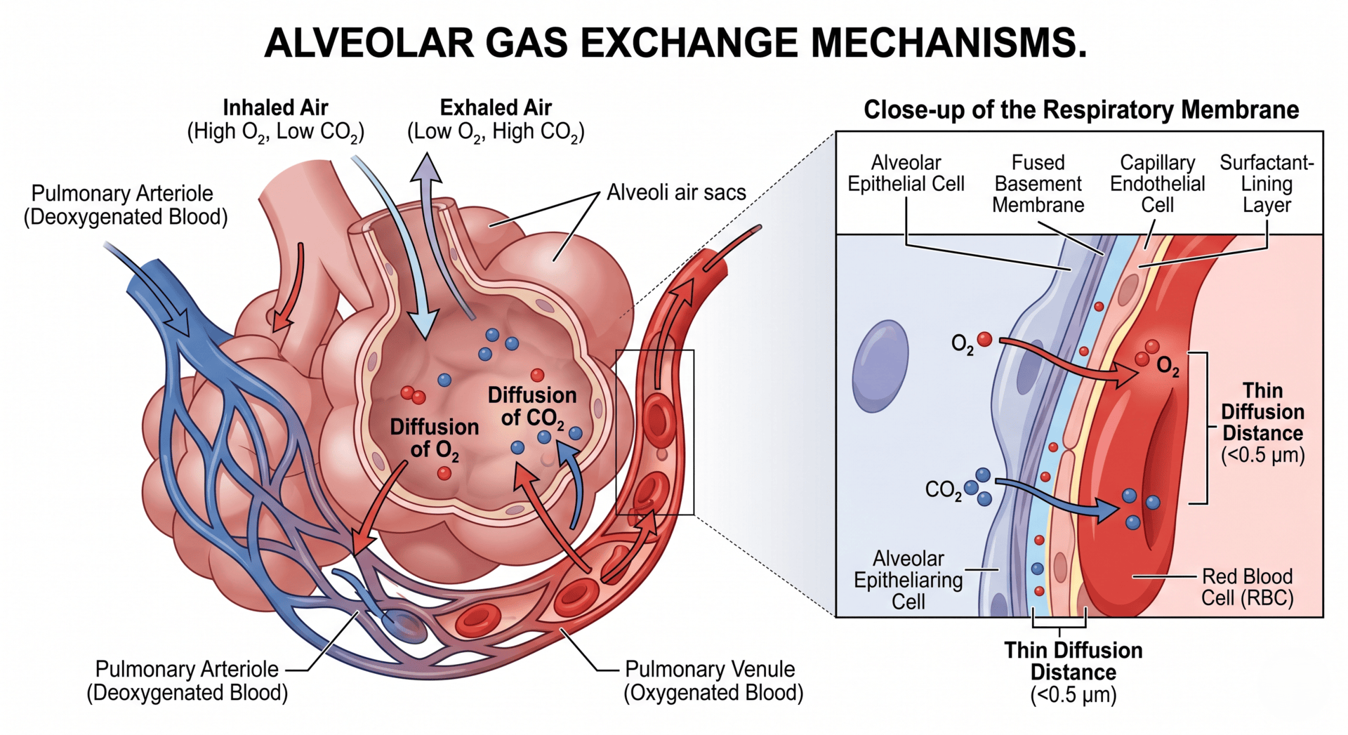
Core Adaptations of the Alveoli
To maximise the movement of molecules via passive diffusion, the alveoli possess specific structural specialisations. These properties directly fulfil the biological requirements outlined by Fick's Law of Diffusion:
1. Large Total Surface Area
The human lungs contain roughly 300 to 500 million alveoli. This massive collective clustering expands the total surface area available for diffusion to approximately 70 square metres (roughly the size of half a tennis court). A larger surface area allows a greater number of gas molecules to cross the membrane simultaneously.
2. Microscopic Diffusion Distance
The barrier between the air in the alveolus and the red blood cells in the capillary is incredibly thin, typically measuring less than 0.5 micrometres. This short diffusion pathway is achieved via two main features:
- The alveolar wall consists of a single layer of flat squamous epithelial cells.
- The capillary wall consists of a single layer of endothelial cells.
3. Rich Capillary Network (Extensive Blood Supply)
Each alveolus is wrapped in a dense mesh of capillaries supplied by the pulmonary artery. This continuous flow of blood instantly whisks oxygenated blood away from the lungs and constantly delivers deoxygenated blood rich in carbon dioxide. This constant movement preserves a steep concentration gradient.
4. Moist Alveolar Lining
The internal surface of the alveoli is lined with a thin film of moisture. Oxygen dissolves in this fluid layer before diffusing across the epithelial membrane. Dissolved gases move across biological bilayers much more rapidly than gases in a purely gaseous phase.
5. Ventilation Mechanisms
Continuous breathing (inspiration and expiration) ensures that stale air low in oxygen and high in carbon dioxide is regularly replaced with fresh atmospheric air. This keeps the concentration of oxygen inside the alveolus higher than that in the incoming blood, driving inward diffusion.
Inspiration vs. Expiration
Ventilation relies on altering the volume of the thoracic cavity to generate pressure gradients between the lungs and the atmosphere.
Inspiration (Breathing In)
- The external intercostal muscles contract, pulling the ribcage upwards and outwards.
- The diaphragm contracts and flattens downward.
- These movements increase the volume of the thoracic cavity, causing a drop in internal air pressure below atmospheric pressure.
- Air is drawn into the lungs down a pressure gradient.
Expiration (Breathing Out)
- The external intercostal muscles relax, causing the ribcage to drop downwards and inwards under its own weight.
- The diaphragm relaxes and domes upward into the chest cavity.
- These movements reduce the volume of the thoracic cavity, raising internal air pressure above atmospheric pressure.
- Air is forced out of the respiratory tract.
Comparative Gas Composition
During gas exchange, oxygen is extracted from the air while carbon dioxide is dumped into it. The differences between inspired and expired air reveal the net efficacy of this system.
The following data outline the exact gaseous shifts observed between inhaled and exhaled air under resting conditions:
| Gas | Inhaled Air (%) | Exhaled Air (%) |
|---|---|---|
| Nitrogen | 78 | 79 |
| Oxygen | 21 | 16 |
| Carbon Dioxide | 0.04 | 4.00 |
| Other Gases (mostly Argon) | 1 | 1 |
Note on Nitrogen: The apparent increase in nitrogen percentage from 78% to 79% is not due to the body creating nitrogen. Because the absolute volume of oxygen drops and water vapour increases in exhaled air, the relative ratio of nitrogen shifts slightly upward in the final mixture.
Worked Example
A student is asked to analyse the mathematical efficiency of gas exchange by calculating the percentage change in carbon dioxide concentration between inhaled and exhaled air using the values provided in the data set.
Step 1: Identify the initial and final values
- Inhaled Carbon Dioxide (Initial Value) = 0.04%
- Exhaled Carbon Dioxide (Final Value) = 4.00%
Step 2: Set up the percentage change formula
To find the relative increase, use the standard percentage change expression:
Step 3: Substitute the values and compute
Subtract the initial concentration from the final concentration:
Divide the difference by the initial concentration:
Multiply by 100 to convert to a percentage:
Summarise with AI:
Did you like this article? Rate it!


i like the article
Hi Sam! Thank you! Great to hear that you found this resource useful.
welcome
Hi Suunu! Thanks for reaching out. Hope you’ve found this resource useful 😊!
i have learnt a lot which i did not know
am senior three student but this is cool
Am impressed
Am impressed
learning is the best