In this article, we will discuss how to calculate the actual size of the specimens from photomicrographs, how to use an eyepiece graticule and stage micrometer scale to make measurements, and differences between resolution and magnification. Besides this, we will also compare electron and light microscopes. So, let us get started.


Actual Size of the Specimen
Example: Computing the Actual Size of the Specimen
Suppose that a scientist views the sample of the red blood cells under a light microscope.
The magnification of the eyepiece lens is x10 and the object lens is x40. A person takes the image of the blood cells from a light microscope (photomicrograph) and he finds out that the average cell size is 5 mm.
Using the above information, your task is to calculate the average size of the red blood cells in the sample. Write the final answer in micrometers.
Given values:
Magnification of an eyepiece lens: x10
Magnification of an objective lens: x40
Size of an image: 3 mm
Follow the steps below to calculate the specimen size.
Step 1: Compute the total magnification of the specimen
The formula for total magnification is given below:
Total magnification = eyepiece lens magnification x Objective lens magnification
= x10 x x40 = x400
Step 2: Compute the size of the image in micrometers
1 mm = 1000 micrometers
5 mm = 5000 micrometers
Step 3: Compute the actual size of the red blood cell
Use the formula below to calculate the actual size of the red blood cell:
Actual size = 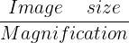
= 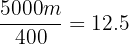 μm
μm
Hence, the average size of the red blood cell in the sample is 12.5 micrometers.
What are Eyepiece Graticules and Stage Micrometers?
To calculate the size of an object as observed under a microscope, we use an eyepiece graticule and a stage micrometer.
We know that every microscope is different so when using there is a need for calibration. A stage micrometer is used to perform this calibration. A stage micrometer is a slide that contains a perfectly accurate scale in micrometers. It generally has 10 m divisions and 1 mm has 100 divisions.
The eyepiece graticule refers to the disc placed in the eyepiece that has 100 divisions. The eyepiece graticule does not contain any scale. To determine the divisions that are equal at each magnification, an eyepiece graticule is calibrated to the stage micrometer at each magnification.
How to use a stage micrometer and an eyepiece graticule?
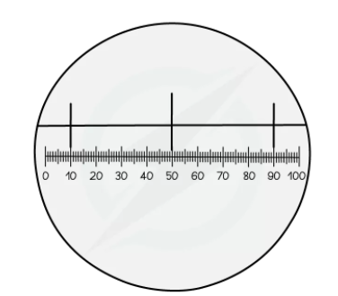
- In the above figure, the stage micrometer contains three lines.
- The distance between each line is 10 μm
- Each 10 μm division contains 40 eyepiece graticule divisions
- 40 graticule divisions are equal to 10 μm
Remember the following formula:
1 graticule division = 
1 graticule division =  μm
μm
0.25 μm is the magnification factor.
- A specimen slide will be used instead of the stage micrometer and the eyepiece graticule at the same magnification to calculate the length of an object.
- Multiply the number of graticule divisions with the magnification factor
Magnification factor x graticule divisions = measurement (in μm)
In the next section of the article, we will discuss resolution and magnification.
Magnification and Resolution
Magnification
Magnification refers to the number of times an image of the specimen observed is bigger than the actual size of the specimen
There are two types of lens in a light microscope:
- An eyepiece lens having the magnification of x10
- A series of (mostly three) objective lenses. The magnification of each lens varies.
To determine the total magnification, multiply the magnification of an eyepiece lens the objective lens.
Total magnification = Magnification of an eyepiece lens x magnification of an objective lens
Resolution
Resolution refers to the ability to differentiate between two different points
- If it is not possible to resolve two distinct points, then they will be viewed as a single point
- The resolution of the light microscope is restricted to the wavelength of light
- Light will be diffracted as it passes through the specimen
- The diffraction of the light depends on its wavelength, i.e. the longer is the wavelength of the light, the more it will be diffracted. As the points get near to each other, the diffraction will overlap
- The resolution of electron microscopes is higher than that of the light microscope because electrons have a smaller wavelength than that of visible light. It means that they can be quite nearer before the overlapping of diffracted beams
- Due to the concept of resolution, the phospholipid bilayer structure of the cell membrane cannot be viewed under a light microscope. Phospholipid bilayer width is about 10 nm and the highest resolution of the light microscope is 200 nm. If the distance between any points is less than 200 nm, then they cannot be resolved by a light microscope and hence cannot be differentiated as separate.
In the next section of the article, we will compare electron and light microscopes.
Comparing Electron and Light Microscope
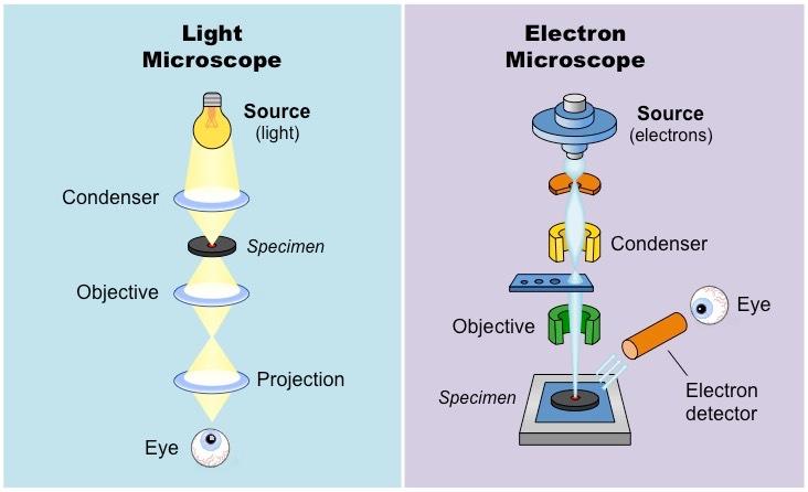
Light Microscopes
- For specimens above 200 nm, we use light microscopes
- Under light microscopes, the light is shined through the specimen, and the light is passed both through an objective lens and an eyepiece lens (x10). Both the lenses magnify the specimen so that it can be seen with the naked eye
- The specimens can either be living or dead
- We can use light microscopes to view the entire cells, small plant, and animal organisms, and tissues inside the organs, for instance, skin or leaves
Electron Microscopes
- Electron microscopes (scanning and transmission) are employed for specimens that are above 0.5 nm
- A beam of electrons is fired by the electron microscopes at the specimen. This beam of light can be either transmission or scanning.
- Transmission is a broad static beam and scanning is a small beam that moves across the specimen.
- An electromagnetic lens picks up the electrons which then shows the image
- Since electron waves are characterized by their higher frequency as compared to visible light, therefore the magnification and resolution of an electron microscope are superior to that of a light microscope.
- We use electron microscopes to observe viruses, organelles, and DNA. Moreover, they are also used to observe the entire cell in more detail.
- The specimen should be dead for viewing under an electron microscope.
Summarise with AI:
Did you like this article? Rate it!