In this article, we will discuss arteries, veins, and capillaries in detail. So, let us get started.


Introduction
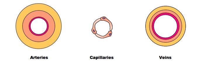
Arteries, veins, and capillaries have different characteristics which show that they play distinctive roles throughout the body. The walls of the arteries and veins are composed of the same component; however, the component is present in varying proportions. Besides this, the wall thickness of the arteries and veins also varies. Now, let us discuss arteries, veins, and capillaries one by one in detail.
Arteries
The blood vessels that carry blood at high pressures away from the heart are known as arteries. They have relatively thicker walls which enable them to tolerate high blood pressure as it rises through every ventricular contraction of the heart. The walls of the arteries are made up of elastic and muscular tissue, and collagen fibres. Arteries that are present near the heart have a higher proportion of elastic fibres. The walls of these arteries must stretch and recoil to accommodate the rising blood pressure. It prevents them from bursting or from dropping blood pressure. These arteries are termed elastic.
The arteries away from the heart have less elastic and more smooth muscle tissue. The diameter of these arteries can be modified to change the flow of blood to various tissues. These arteries are considered as being muscular and they have small branches known as arterioles.
The lumen of the arteries is comparatively lower to guarantee that blood remains at relatively high pressure for efficient delivery to the tissues. It also provides resistance to the flow of blood to enable the exchange of has as blood passes through the tissues.
Capillaries
Arterioles divide into smaller branches known as capillaries. The capillaries form networks throughout the majority of the tissues in the body. The diameter of the capillaries ranges from 5 to 10 μm.
Blood that flows through the capillaries is brought near to the cells of the body to enable the efficient exchange of materials. The one-cell thick endothelial walls of the capillaries guarantee the easy diffusion of substances between the capillaries and the adjacent cells. There are tiny gaps between individual squamous epithelial cells that create the wall to enable the leakage of tiny substances out of the blood into the fluid surrounding the cells of the body.
Veins
Capillaries combine to create larger blood vessels known as venules which join to form veins. Veins have a relatively tough outer layer that is made up of collagen fibres. On the other hand, the intermediate layer is comparatively thin and has only a tiny amount of elastic fibre and smooth muscle.
Veins have a characteristically large lumen. Skeletal muscle contraction assists in increasing the blood pressure temporarily within the veins. Moreover, one-way valves prevent the blood from flowing back toward the heart.
In the next section, we will explain how the structure of muscular arteries, elastic arteries, veins, and capillaries is related to their functions.
Structure and Related Functions of Muscular Arteries, Elastic Arteries, Veins and Capillaries
Muscular Artery
Structure: Muscular arteries have thinner tunica media which is primarily made up of smooth muscle.
Function: Due to this, muscular arteries can perform vasoconstriction and vasodilation.
Structure: Muscular arteries are very less elastic as compared to elastic arteries.
Function: This structure prevents these arteries from stretching and recoiling.
Structure: Muscular arteries have a narrow lumen
Function: This structure allows the blood to flow under high pressure
Elastic Artery
Structure: Elastic arteries have a thicker tunica media that is primarily made up of elastin and collagen.
Function: This structure allows the elastic artery to stretch in response to each pulse
Structure: Elastic arteries have comparatively less smooth muscle fibres
Function: This structure prevents these arteries from performing vasoconstriction and vasodilation.
Structure: Elastic arteries have a narrow lumen
Function: This structure enables the blood to flow under high pressure
Veins
Structure: One-way valves are present
Function: Prevents the backward flow of blood
Structure: Veins have a wide lumen
Function: This decreases blood pressure in veins with no surges
Structure: Veins have less smooth muscle and elastin
Function: It prevents the veins from stretching and recoiling
Structure: Veins have a lot of collagen
Function: It enhances their strength and structure
Capillary
Structure: Capillaries have a very small diameter
Function: Blood moves comparatively slower, giving more opportunity for diffusion to occur
Structure: They branch between the cells
Function: This structure allows the substances to diffuse quickly between the cells
Structure: Capillaries have thin walls. There is no elastin, smooth muscle, or collagen.
Function: This structure allows them to fit between individual cells and diffusion becomes quicker.
In the next section of the article, we will discuss and draw red blood cells, monocytes, neutrophils, and lymphocytes.
Blood Cells
Blood is a tissue that is made up of many important specialized cells. Monocytes, red blood cells, neutrophils, and lymphocytes have distinctive features that allow them to be recognized in photomicrographs, on microscopic slides, and in electron micrographs.
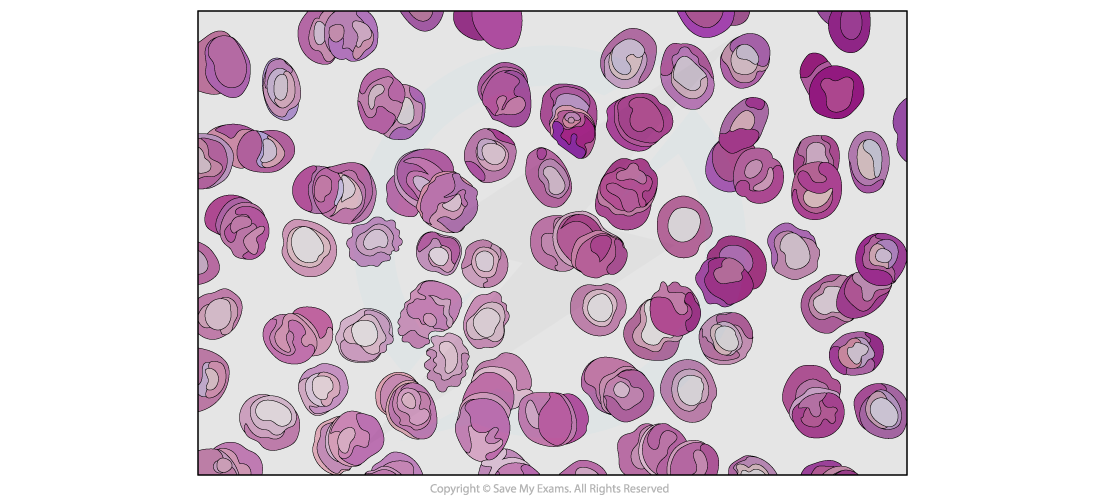
Red blood cells
There are almost 5 million red blood cells per mm3 of blood. Red blood cells have haemoglobin which is a protein with a quaternary structure that has haem iron groups that can reversibly bind to oxygen.
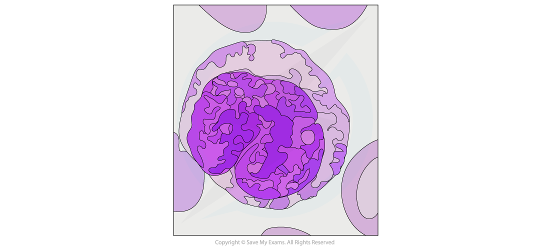
Monocytes
We can recognize monocytes by their size as they are the largest of the leukocytes and contain a nucleus that resembles a bean or a kidney. The nucleus of the monocytes tends to be lighter when viewed after staining as compared to other leukocytes. The nucleus should be light blue, whereas the chromatin inside is fine and distinct.
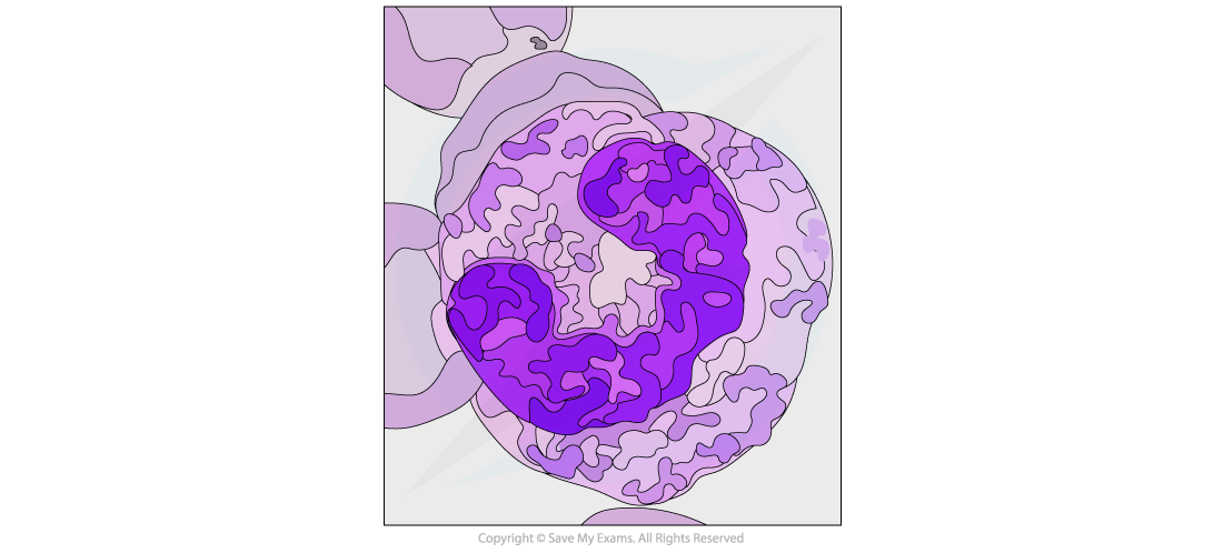
Neutrophils
The neutrophils can be identified by their multi0lobed nuclei. Approximately 70% of all leukocytes are neutrophils which makes them easy to be viewed on a micrograph. The granules of the neutrophils generally stain purple-blue or pink.
Lymphocytes
Lymphocytes refer to the tiny leukocytes that are distinguishable by their extremely large nuclei that generally stain a dark color. Approximately 20 to 25% of all leukocytes are lymphocytes. The size of the lymphocytes and the red blood cells is almost the same.

Summarise with AI:
Did you like this article? Rate it!