At A Level Biology, you are expected to go well beyond the GCSE comparison of four basic components. You must be able to describe the ultrastructure of plant and animal cells as seen in electron micrographs, explain the function of every organelle with precision, account for structural differences in terms of the different metabolic demands of the two cell types, and apply this knowledge to unfamiliar specialised cells in exam questions. This article covers all of these requirements.
Prerequisite: The GCSE article Structure of Plant and Animal Cells covers the basic light-microscope comparison. This article assumes that knowledge and builds from it.


1. Eukaryotic Cells: The Shared Framework
Both plant and animal cells are eukaryotic. The defining feature of a eukaryotic cell is that its genetic material is enclosed within a membrane-bound nucleus. Eukaryotic cells also contain a range of membrane-bound organelles, each maintaining a distinct internal environment optimised for a specific function. This compartmentalisation allows incompatible reactions to occur simultaneously within the same cell.
The organelles common to both cell types are described below with the level of structural detail required at A-Level.
The Nucleus
The nucleus is typically spherical and 10–20 μm in diameter — large enough to be seen with a light microscope. It is bounded by the nuclear envelope, a double membrane whose outer layer is continuous with the rough endoplasmic reticulum. The envelope is perforated by nuclear pores, each 40–100 nm in diameter, through which mRNA, ribosomal subunits, and regulatory proteins can pass. The interior contains nucleoplasm in which chromatin (DNA tightly coiled around histone proteins) is suspended. One or more nucleoli are visible within the nucleus; the nucleolus is the site of ribosomal RNA (rRNA) synthesis and ribosome assembly.
• Nuclear envelope (double membrane) — isolates DNA from cytoplasm, maintaining distinct environments for transcription and translation.
• Nuclear pores — selective passage of mRNA out (for translation at ribosomes) and enzymes in (e.g. DNA polymerase for replication).
• Chromatin/histones — DNA compaction; histones also regulate gene expression by controlling access to DNA.
• Nucleolus — ribosome production; cells with high rates of protein synthesis (e.g. secretory cells) have large, prominent nucleoli.
Mitochondria
Mitochondria are rod-shaped organelles, 1–10 μm in length, enclosed by a double membrane. The outer membrane is smooth; the inner membrane is extensively folded into finger-like projections called cristae, which greatly increase the surface area available for the proteins of the electron transport chain. The internal space enclosed by the inner membrane is the matrix, a dense fluid containing the enzymes of the Krebs cycle, circular mitochondrial DNA (mtDNA), and 70S ribosomes. The presence of their own DNA and 70S (not 80S) ribosomes is evidence for the endosymbiotic origin of mitochondria from ancient prokaryotes.
Cells with high energy demands (e.g. muscle fibres, epithelial cells of the ileum, liver cells) contain large numbers of mitochondria with densely packed cristae. In exam questions, always link 'many mitochondria' explicitly to 'high rate of aerobic respiration' and 'ATP production', not simply to 'energy'.
Ribosomes
Ribosomes are non-membrane-bound organelles, approximately 20–25 nm in diameter, each comprising a large and a small subunit made of ribosomal RNA and protein. In eukaryotic cells, cytoplasmic ribosomes are 80S (composed of 60S and 40S subunits). They occur either free in the cytoplasm (synthesising proteins for use within the cell) or bound to the rough endoplasmic reticulum (synthesising proteins destined for secretion or membrane insertion).
Mitochondria and chloroplasts contain their own ribosomes, which are 70S — the same size as prokaryotic ribosomes. This is a frequently tested distinction. '70S ribosomes' is the correct answer if a question asks what organelle feature supports the endosymbiotic theory.
Endoplasmic Reticulum (ER)
The endoplasmic reticulum is a continuous network of membrane-bound cisternae (flattened sacs) that extends throughout the cytoplasm and is connected to the outer nuclear membrane.
| Type | Key structural feature | Primary functions |
|---|---|---|
| Rough ER (RER) | Ribosomes studded on outer surface | Synthesis of proteins destined for secretion, membrane insertion, or the Golgi apparatus; folds and glycosylates polypeptide chains |
| Smooth ER (SER) | No ribosomes | Synthesis of lipids and phospholipids; steroid synthesis; detoxification of drugs and metabolites; stores calcium ions in muscle cells |
Golgi Apparatus
The Golgi apparatus consists of a stack of flattened, membrane-bound cisternae with a characteristic curved, 'wifi-symbol' profile in electron micrographs. Proteins and lipids arriving from the RER enter the cis face (nearest to the ER) and travel through the stack by vesicle budding and fusion, undergoing progressive modification — glycosylation, phosphorylation, and sorting — before leaving from the trans face in vesicles. These vesicles may fuse with the plasma membrane for secretion (exocytosis), remain in the cytoplasm as lysosomes, or be directed to specific organelles.
Secretory pathway summary: Ribosome → RER → Golgi apparatus → secretory vesicle → plasma membrane (exocytosis). This pathway is the route for all extracellular proteins, including digestive enzymes, antibodies, and hormones such as insulin.
Lysosomes
Lysosomes are spherical, membrane-bound vesicles, 0.1–0.5 μm in diameter, produced by the Golgi apparatus. They contain a variety of hydrolytic enzymes (lysozymes) maintained at an acidic pH (~4.5–5.0) by proton pumps in the lysosomal membrane. Lysosomes function in:
- Autophagy — digesting worn-out organelles whose components can be recycled.
- Phagocytosis — fusing with phagosomes to digest engulfed pathogens (key function in macrophages and neutrophils).
- Apoptosis — releasing enzymes to facilitate controlled cell death.
Animal cells contain prominent lysosomes; they are less prominent in plant cells. The plant vacuole can perform a similar digestive role. If a question describes a cell with 'many membrane-bound vesicles containing digestive enzymes', it is describing a cell rich in lysosomes — typically a phagocyte.
Centrioles (Animal Cells Only)
Centrioles are short, cylindrical structures (~0.3 μm long) composed of nine triplets of microtubules arranged in a ring, with no central pair. Two centrioles arranged at right angles to each other form a centrosome. During cell division, the centrosome organises the mitotic spindle by nucleating spindle fibres (microtubules) that attach to chromosomes at the kinetochore. Centrioles are absent from higher plant cells, which use alternative mechanisms to organise their spindle.
Note: This is a key difference between plant and animal cells often tested in A-Level questions. The statement 'centrioles are present in animal cells but absent in most plant cells' must be supported by the functional explanation that animals use centrioles to organise the spindle during mitosis.
Microvilli (Specialised Animal Cell Feature)
Microvilli are finger-like extensions of the plasma membrane, 1–3 μm long, found on the apical surfaces of epithelial cells lining the small intestine (where they form the brush border) and renal tubules. Each microvillus contains a core of actin filaments anchored to the cortical cytoskeleton. Microvilli massively increase the surface area of the cell surface membrane, enhancing the rate of absorption by diffusion and co-transport.
2. Organelles Found Only in Plant Cells
Cell Wall
The plant cell wall lies outside the plasma membrane and is a non-living structure made primarily of cellulose microfibrils embedded in a matrix of hemicellulose, pectin, and glycoproteins. Cellulose is a polysaccharide of β-glucose monomers linked by 1,4-glycosidic bonds, which gives it a straight, unbranched structure that allows hydrogen bonding between parallel chains to form strong microfibrils. The cell wall is freely permeable to water and dissolved solutes, unlike the selectively permeable plasma membrane.
The primary cell wall is laid down first; some cells (e.g. xylem vessels) subsequently deposit a thicker, lignified secondary cell wall. Cell walls of adjacent cells are joined by the middle lamella, composed of calcium pectate.
| Function | Structural basis |
|---|---|
| Provides mechanical support and prevents over-expansion | Rigidity of cellulose microfibrils; turgor pressure acts against the inextensible wall |
| Maintains cell shape | Inflexible lattice of cellulose |
| Allows turgor pressure to develop | Wall withstands pressure of water drawn in by osmosis |
| Cell-to-cell communication via plasmodesmata | Pores traversing the wall filled with cytoplasmic strands |
Plasmodesmata
Plasmodesmata (singular: plasmodesma) are narrow channels, approximately 50–60 nm in diameter, that pass through the cell walls of adjacent plant cells. Each channel is lined with plasma membrane and contains a central strand of smooth ER called the desmotubule. Plasmodesmata allow direct cytoplasmic continuity between neighbouring cells — the connected cytoplasm is called the symplast. They permit the movement of water, ions, sugars (e.g. sucrose from mesophyll cells into phloem), signalling molecules, and even viral RNA between cells, without molecules having to cross a membrane.
The symplast pathway (through plasmodesmata) and the apoplast pathway (through cell walls, not crossing membranes) are both required knowledge for A-Level. Be clear that plasmodesmata are the structural basis of symplastic transport.
Chloroplasts
Chloroplasts are large biconvex organelles, 4–10 μm long and 2–3 μm wide, found primarily in the mesophyll cells of leaves. Like mitochondria, they are bounded by a double membrane (the chloroplast envelope), possess their own circular DNA and 70S ribosomes, and show evidence of endosymbiotic origin from photosynthetic prokaryotes.
The internal membrane system comprises thylakoids — flattened, membrane-bound sacs stacked into grana (singular: granum). Individual grana are connected by intergranal lamellae (stromal lamellae). The fluid surrounding the thylakoids is the stroma.
| Compartment | Location of photosynthesis stage | Key components |
|---|---|---|
| Thylakoid membranes / grana | Light-dependent reactions | Photosystems I and II; electron transport chain; ATP synthase; chlorophyll and carotenoid pigments |
| Stroma | Light-independent reactions (Calvin cycle) | RuBisCO; NADPH; ATP; 70S ribosomes; circular chloroplast DNA |
Chloroplasts contain their own ribosomes (70S) and circular DNA, enabling them to synthesise some of their own proteins. However, the majority of chloroplast proteins are encoded by nuclear DNA and imported post-translationally.
Permanent Central Vacuole and Tonoplast
Mature plant cells typically contain a single large central vacuole that can occupy up to 90% of the cell volume. It is bounded by the tonoplast, a selectively permeable membrane. The vacuole contains cell sap — an aqueous solution of sugars, ions, pigments (anthocyanins in many flowers and fruits), and organic acids. Key functions include:
- Maintaining turgor pressure: water enters the vacuole by osmosis when the surrounding solution is more dilute than the cell sap, pushing the plasma membrane against the cell wall (turgid state). This provides structural support to non-woody tissues such as leaves and young stems.
- Storage: of sugars, ions (e.g. nitrate, potassium), pigments, and waste products (e.g. oxalate crystals, tannins).
- Lytic function: the vacuole in some plant cells contains hydrolases and acts like a large lysosome, breaking down cellular debris.
In contrast, animal cells may contain small, temporary vacuoles (e.g. food vacuoles in phagocytes) but lack a large permanent central vacuole.
3. The Plasma Membrane (Cell Surface Membrane)
All cells are bounded by a plasma membrane, which in plant cells lies inside the cell wall. The currently accepted model of membrane structure is the fluid mosaic model, proposed by Singer and Nicolson in 1972.
The Fluid Mosaic Model
'Fluid' refers to the lateral mobility of phospholipid molecules and many membrane proteins within the bilayer at physiological temperatures. 'Mosaic' refers to the diverse assortment of proteins embedded within and on the surface of the bilayer.
| Component | Structure | Function |
|---|---|---|
| Phospholipid bilayer | Hydrophilic phosphate heads face outwards (towards aqueous environments); hydrophobic fatty acid tails face inwards | Forms the basic barrier; hydrophobic core prevents free passage of polar and charged molecules |
| Cholesterol | A sterol molecule that sits among the fatty acid tails of the bilayer | Reduces fluidity at high temperatures (prevents excessive lateral movement); prevents crystallisation at low temperatures — stabilises membrane fluidity over a range of temperatures. Not found in prokaryotic membranes. |
| Intrinsic (integral) proteins | Span the entire bilayer; include channel proteins and carrier proteins | Channel proteins: aqueous pores for facilitated diffusion of ions and polar molecules. Carrier proteins: bind specific molecules and change conformation to transport them (facilitated diffusion and active transport). |
| Extrinsic (peripheral) proteins | Attached to the surface of the bilayer, not spanning it | Cell signalling; mechanical support; linked to glycoproteins and glycolipids for cell recognition |
| Glycoproteins | Proteins with carbohydrate chains on the extracellular face | Cell recognition (e.g. ABO blood group antigens); receptor binding for hormones and neurotransmitters; cell adhesion |
| Glycolipids | Phospholipids with carbohydrate chains on the extracellular face | Cell recognition; stabilising membrane structure |
Examiners frequently ask you to 'explain why the membrane is described as fluid' — the answer is lateral movement of phospholipids. Examiners also ask why cholesterol is important — link it to temperature: prevents the membrane becoming too fluid at high temperature OR too rigid (crystalline) at low temperature.
4. Direct Comparison: Plant vs Animal Cell Ultrastructure
| Feature | Animal cell | Plant cell |
|---|---|---|
| Plasma membrane | Present — outermost boundary | Present — inside cell wall |
| Cell wall | Absent | Present — cellulose microfibrils; provides rigidity and turgor support |
| Nucleus | Present — central position; chromatin + nucleolus | Present — peripheral position (pushed aside by vacuole); chromatin + nucleolus |
| Mitochondria | Present (many in active cells) | Present (many in active cells) |
| Chloroplasts | Absent | Present in photosynthetic cells (e.g. mesophyll); contain thylakoids, grana, stroma |
| Rough ER | Present | Present |
| Smooth ER | Present | Present |
| Golgi apparatus | Present | Present |
| Ribosomes (80S) | Present — free and on RER | Present — free and on RER |
| Lysosomes | Present — prominent; key role in phagocytosis | Less prominent; vacuole performs some lytic functions |
| Centrioles | Present — organise mitotic spindle | Absent in higher plant cells; spindle organised differently |
| Permanent large vacuole | Absent (only small, temporary vacuoles) | Present — central; bounded by tonoplast; maintains turgor |
| Plasmodesmata | Absent | Present — cytoplasmic channels through cell walls; allow symplastic transport |
| Microvilli | Present on some cells (e.g. ileum epithelium, renal tubule) | Absent |
| Cell shape | Irregular; variable (no rigid wall) | Regular; often rectangular in cross-section (constrained by rigid wall) |
| Starch grains | Absent | Present in chloroplasts and amyloplasts |
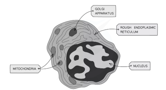
5. Electron Micrographs of Plant and Animal Cells
A-Level exams regularly present transmission electron micrograph (TEM) images and ask you to identify organelles and explain their functions. The following features identify each organelle:
| Organelle | Identifying features in a TEM |
|---|---|
| Nucleus | Large, dark-staining; double membrane (nuclear envelope visible as two parallel lines); nuclear pores; darker nucleolus within |
| Mitochondrion | Oval/rod-shaped; double membrane; inner membrane folded into cristae (appear as finger-like projections or shelf-like folds in cross-section); dense matrix |
| Chloroplast | Larger than mitochondrion; double envelope; internal stacks of thylakoids (grana appear as dark parallel lines); lighter stroma surrounding them |
| Rough ER | Network of membrane-bound cisternae; ribosomes visible as dark dots on outer surface; connected to nuclear envelope |
| Smooth ER | Tubular membrane network; no ribosomes on surface |
| Golgi apparatus | Stack of flattened cisternae, curved at edges; vesicles budding from trans face |
| Lysosome | Small, spherical, membrane-bound; uniform dense contents |
| Ribosome | Tiny dark dots (~20 nm); found free in cytoplasm or on RER surface |
| Centriole | Two short cylinders at right angles; 9 triplets of microtubules (seen in cross-section as a ring of 9 'C'-shaped profiles) |
| Cell wall (plant) | Thick, electron-dense layer outside plasma membrane; no membrane of its own |
| Vacuole (plant) | Large, light-staining area; single tonoplast membrane visible |
| Plasmodesmata (plant) | Fine channels crossing the cell wall, connecting adjacent cells; visible as narrow dark lines |
An electron micrograph refers to the image of a specimen taken using an electron microscope.
The electron micrograph of an animal cell is shown below:
The electron micrograph of a plant cell is shown below:

Now, what do these electron micrographs of both the cells indicate?
Applying Ultrastructure to Specialised Cells
A-Level questions often describe an unfamiliar specialised cell and ask you to explain its features in terms of organelle ultrastructure. The key skill is linking the increased quantity or size of an organelle to the specific function it serves.
| Specialised cell | Notable ultrastructural features | Functional explanation |
|---|---|---|
| Ileum epithelial cell (enterocyte) | Many microvilli (brush border); abundant mitochondria; extensive RER and Golgi | Microvilli increase SA for absorption; mitochondria provide ATP for active transport and co-transport; RER/Golgi produce secreted enzymes (e.g. maltase) |
| Pancreatic acinar cell | Very large nucleolus; extensive RER; prominent Golgi; many secretory vesicles | Produces and secretes large quantities of digestive enzymes (proteins) — requires high ribosome production, synthesis, processing and packaging |
| Muscle fibre (myocyte) | Numerous large mitochondria with many cristae | High ATP demand for myosin cross-bridge cycling during contraction |
| Palisade mesophyll cell (plant) | Many chloroplasts positioned parallel to upper leaf surface; large vacuole | Chloroplasts maximise light absorption for photosynthesis; vacuole maintains turgor to keep cells and leaf rigid |
| Guard cell (plant) | Chloroplasts present; large vacuole; distinctive cell wall thickening | Chloroplasts produce ATP and sugar (involved in opening mechanism); vacuole changes in volume to control stomatal aperture |
| Neutrophil (white blood cell) | Multi-lobed nucleus; many lysosomes; no chloroplasts or cell wall | Lobed nucleus allows squeezing through capillary walls (diapedesis); lysosomes contain enzymes to destroy engulfed pathogens after phagocytosis |
Well, we can conclude the following two points from these electron micrographs:
- Centrioles and microvilli are present in animal cells but absent in plant cells.
- Additional structures such as cell walls made up of cellulose, chloroplasts, and large permanent vacuoles are present in plant cells but absent in animal cells.
- The structure of plant cells is larger and more regular as compared to the structure of an animal cell.
Summarise with AI:
Did you like this article? Rate it!