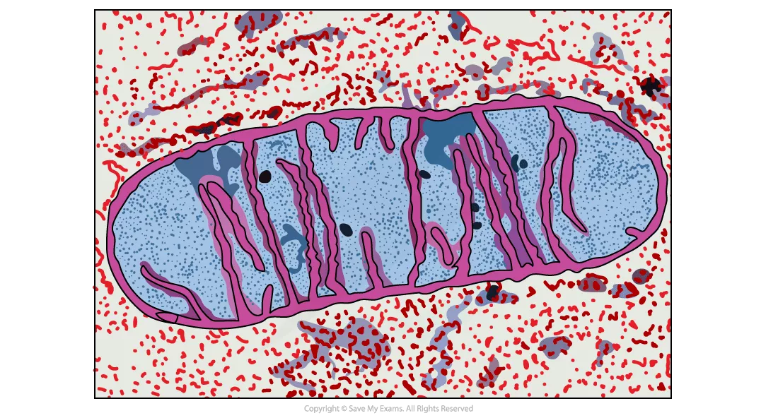
In this article, we will discuss the structure, function, and relationship between the structure and function of mitochondria. To understand the structure of mitochondria, we will use diagrams and electron micrographs. So, let us get started.


Introduction
Mitochondria refer to rod-shaped organelles which are usually 0.5 to 1 micrometre in diameter. They are bacteria-sized organelles and are widely found in most eukaryotic cells. Generally, there are almost 2000 mitochondria in a single cell. It means that the mitochondria represent almost 25% of the cell volume.
In eukaryotic cells, mitochondria are the site of aerobic respiration. Aerobic respiration is a type of respiration that occurs in the presence of oxygen. The primary function of mitochondria is ATP synthesis. The ATP synthesis in mitochondria takes place during the final stage of respiration and is known as oxidative phosphorylation. The oxidative phosphorylation depends on membrane proteins that make up the ATP synthase enzyme and electron transport chain.
In the next section of the article, we will discuss the structure of mitochondria in detail.
Structure of Mitochondria
As we already know that mitochondria are small rod-shaped organelles that make up a quarter of the eukaryotic cell’s volume. Although these structures are extremely small, however, they have an elaborate structure that complements the functions they perform.
Membranes
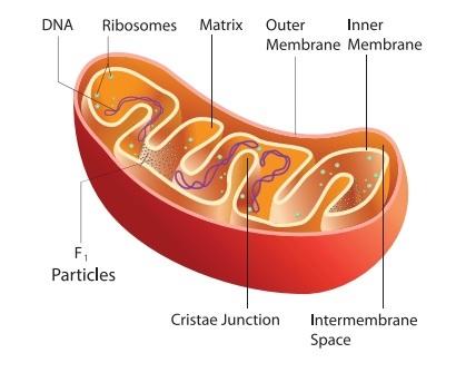
These tiny organelles are enclosed by two phospholipid membranes: a smooth outer membrane and an inner membrane. The inner membrane is markedly folded and entails a large surface and encloses the matrix space.
Besides being smooth, the outer membrane of mitochondria is permeable to many tiny molecules. Permeable means that the outer membrane allows several molecules to pass through it.
The characteristics of the inner membrane are:
- It is less permeable as compared to the outer membrane. It means that it does not allow the molecules to pass through it as much as the outer membrane.
- It is folded and its folds are referred to as cristae. It also entails tubes like protrusions known as tubules.
- It is the site of the electron transport chain employed in oxidative phosphorylation
- It is the location of ATP synthase employed in oxidative phosphorylation
Intermembrane space
Mitochondria also have an intermembrane space. Intermembrane space refers to the space between outer and inner membranes. The main features of intermembrane space are:
- This space entails a low pH because it has a higher concentration of protons.
- The concentration gradient across the inner membrane is created during oxidative phosphorylation and is critical for the synthesis of ATP.
Shape and number of mitochondria
Remember that not only the shape and number but also the number of cristae of the mitochondria vary in different cells. These characteristics depend on the type of the cell.
Tissues that have rigorous oxidative metabolism like heart muscle contain mitochondria that have large numbers of folds known as cristae. The shape of the mitochondria can even differ in a single kind of tissue because these features depend on their functional status.
Development of mitochondria
These mobile, plastic organelles are believed to have developed during an early phase of evolution from aerobic bacteria. These bacteria entered into the symbiosis with primitive anaerobic eukaryotes. Many findings support the endo-symbiont theory. For instance, mitochondria are characterized by their ring-shaped DNA.
Each mitochondrion has four molecules of DNA. Similarly, mitochondria have their own ribosomes. The genome of mitochondria became smaller over the course of evolution.
Mitochondrial envelope
The mitochondrial envelope that has two membranes also supports endosymbiont theory. The inner membrane that is derived from the previous symbiont entails a structure that resembles prokaryotes.
It has distinct lipid cardiolipin, however, it hardly contains any cholesterol. Both mitochondrial membranes contain a lot of proteins. The outer membrane has porins that enable the exchange of tiny molecules between the cytoplasm and intermembrane space.
The inner membrane of mitochondria is impermeable. It does not even allow the smaller molecules to pass through it except oxygen, carbon dioxide, and water. The inner membrane has many transporters that ensure the import and export of critical metabolites. The inner membrane is also responsible for the transport of ATP synthase, respiratory chain complexes, and other enzymes.
Mitochondria in humans
Mitochondria in humans still have 16,569 base pairs. These pairs code for two rRNAs, 22 tRNAs, and 13 proteins. These 13 proteins are only produced in the mitochondrion.
Matrix
The mitochondrion also has a matrix that is rich in enzymes. The matrix of the mitochondria refers to an aqueous solution inside the inner membranes of the mitochondrion. It has enzymes, ribosomes, and circular mitochondrial DNA that are essential for the functioning of mitochondria.
The following diagram shows the structure of mitochondria.
In the next section of the article, we will discuss the functions of mitochondria in detail.
Mitochondria Functions
- The primary function of mitochondria is the production of energy.
- Mitochondria processes the simpler molecules of nutrition and in turn produces charged molecules.
- The combination of these charged molecules with oxygen produces ATP molecules. This process is referred to as oxidative phosphorylation.
- Mitochondria aid the cells to maintain the appropriate concentration of calcium ions with the cell compartments.
- These tiny organelles also assist to build some parts of the blood and hormones, for instance, estrogen and testosterone.
- The mitochondria in liver cells contain enzymes that detoxify ammonia.
- These organelles also play a vital role in the process of apoptosis and programmed death of the cell.
- Unusual cell death because of the dysfunction of mitochondria can impact the function of organs.
In the next section of the article, we will discuss the relationship between the structure and function of mitochondria.
Relationship Between Structure & Function
- Mitochondrion structure is well adapted to the function it performs.
- The larger surface area of the mitochondria because of the inner folds known as cristae helps the membrane to hold several electron transport chain proteins and ATP synthase enzymes.
- Additional active cell types may have larger mitochondria that have longer and tightly packed folds (cristae) so that more ATP can be synthesized.
- The number of mitochondria in each cell differs and depends on the activity of the cell. For example, muscle cells are very active, therefore they contain a large number of mitochondria per cell as compared to fat cells.
Summarise with AI:
Did you like this article? Rate it!