In this article, we will discuss the importance of the refractory period in determining the frequency of impulses. Moreover, we will also explain the structure of a cholinergic synapse and describe how it functions, which will include the role of calcium ions. So, let us get started with the refractory period first.


The Refractory Period
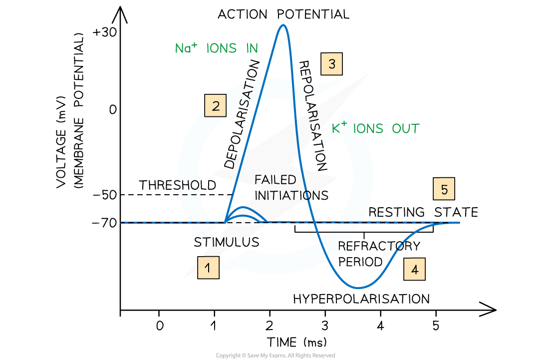
- Soon after the generation of action potential in the part of the axon membrane, all the sodium ion voltage-gated channel proteins in that part or section close. Remember that it happens extremely quickly, approximately 1 ms after the generation of the action potential. It prevents any more sodium ions from diffusing into the axon
- In this part or section of the axon, the potassium ion voltage-gated channel proteins close to enable the diffusion of potassium ions out of the axon, down their concentration gradient
- Due to this, the potential difference slowly returns to normal (approximately -70mV). This process is referred to as repolarization
- The potassium ion voltage-gated channel close and the sodium ion channel proteins in this part of the membrane start responding to depolarization again after the resting potential are near to being established again
- Until this happens, this section or part of the axon membrane is in the recovery period and does not respond, i.e. it becomes unresponsive. This is called the refractory period.
In the next section of the article, we will discuss the importance of the refractory period.
The Importance of Refractory Period
The importance of the refractory period can be established from the following points:
- It prevents the merging of action potentials into each other. In other words, we can say that the refractory period guarantees that action potentials are distinct events
- It guarantees the generation of new action potentials ahead which means further in the axon, instead of behind the original action potential. Remember that the region behind is recovering from the action potential that just took place
- It implies that the impulse can only be transmitted in a single direction only which is critical for transferring nerve impulses along the neurons successfully and efficiently
- It also implies that there is a minimum time between action potentials taking place at a single place along a neuron
- The length of the refractory period is critical for determining the maximum frequency at which the impulses can be transferred along neurons (between 500 to 1000 per second)
In the next section of the article, we will discuss the structure of a cholinergic synapse and describe how it functions, including the role of calcium ions.
Cholinergic Synapses
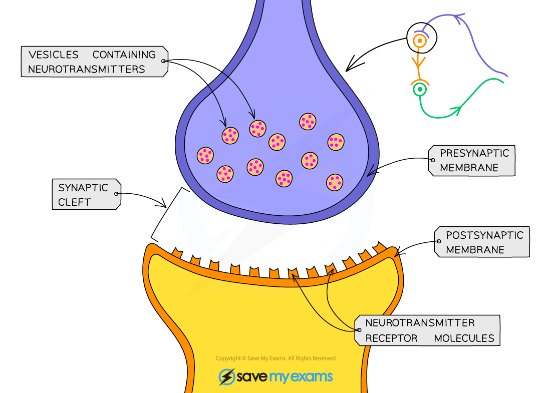
- After the meeting of two neurons, they do not actually come in touch, i.e., physical contact with each other. Instead, an extremely tiny gap, referred to as synaptic cleft, segregates them
- A synapse is created from the ends of the two neurons, along with the synaptic cleft
Let us now discuss the primary mechanism of synaptic transmission.
The Primary Mechanism of the Synaptic Transmission
- Electrical impulses are unable to jump across the synapses
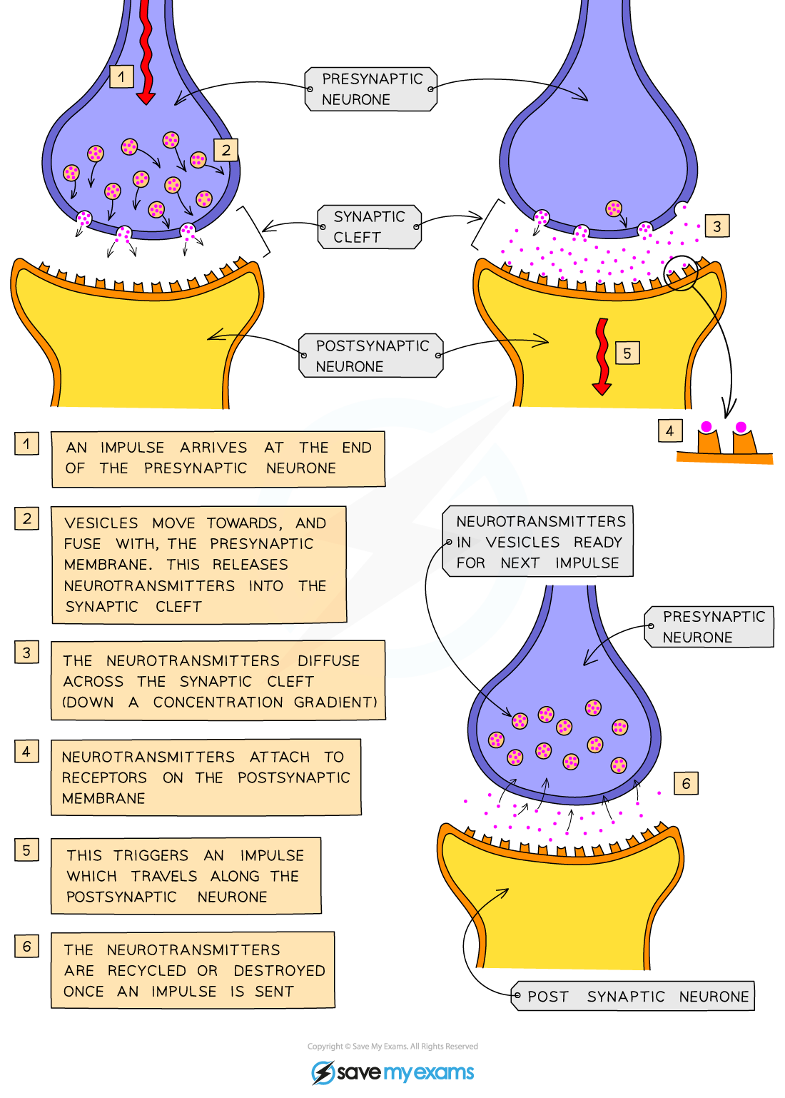
- There is the release of a chemical messenger known as neurotransmitters from vesicles at the presynaptic membrane when an electrical impulse arrives at the end of the axon on the presynaptic neuron
- After that the neurotransmitters that were released earlier diffuse across the synaptic cleft and bind with receptor molecules on the postsynaptic membrane temporarily
- It triggers (stimulates) the postsynaptic neuron so that it can generate an electrical impulse that can be transmitted down the axon of the postsynaptic neuron
- After that, the neurotransmitters are destroyed and recycled, so that any further stimulation of the second neuron can be prevented
- If it is not done, then the repeated impulses can be sent
The Detailed Mechanism of Synaptic Transmission
So far, we have discussed the primary mechanism of synaptic transmission. In this section, we will discuss the detailed mechanism of synaptic transmission.
The total number of known neurotransmitters is 40. Acetylcholine (Ach) is the main neurotransmitter that is employed throughout the nervous system. Cholinergic synapses refer to the synapses that employ the neurotransmitter Ach
Synaptic transmission using ACh takes place using the following process:
- Depolarization of the membrane occurs due to the arrival of an action potential at the presynaptic membrane
- It triggers (stimulates) the opening of voltage-gated calcium ion channel proteins
- Calcium ions diffuse down an electrochemical gradient from the tissue fluid that surrounds the synapse (calcium ions in high concentration) into the cytoplasm of the presynaptic neuron (calcium ions in low concentration)
- Due to this, the ACh-containing vesicles are stimulated to fuse with the presynaptic membrane which releases ACh molecules into the synaptic cleft
- The ACh molecules diffuse across the synaptic cleft and attach (bind) to the receptor proteins in the postsynaptic membrane, however, this binding is temporary
- Because of this, a conformation change in the receptor proteins occurs. The receptor proteins then open to enable the diffusion of sodium ions down an electrochemical gradient into the cytoplasm of the postsynaptic neuron
- The depolarization of the postsynaptic membrane occurs due to the sodium ions, which start the electrical impulse again. This electrical impulse can now continue down the axon of the postsynaptic neuron
- The ACh molecules are broken down and recycled to stop the permanent opening of the sodium ion channels and permanent depolarization of the postsynaptic membrane
- The enzyme known as acetylcholinesterase catalyzes (speeds up) the hydrolysis of ACh molecules into choline and acetate
- The choline gets absorbed again into the presynaptic membrane and forms ACh after it reacts with the acetyl coenzyme A. ACh is then packaged into presynaptic vesicles so that it can be used readily after the arrival of another action potential
- The whole sequence of events occurs in 5 to 10 ms
Summarise with AI:
Did you like this article? Rate it!