In this article, we will discuss the structure of the human gas exchange system in detail. So, let us get started.


Structures Present in Thorax
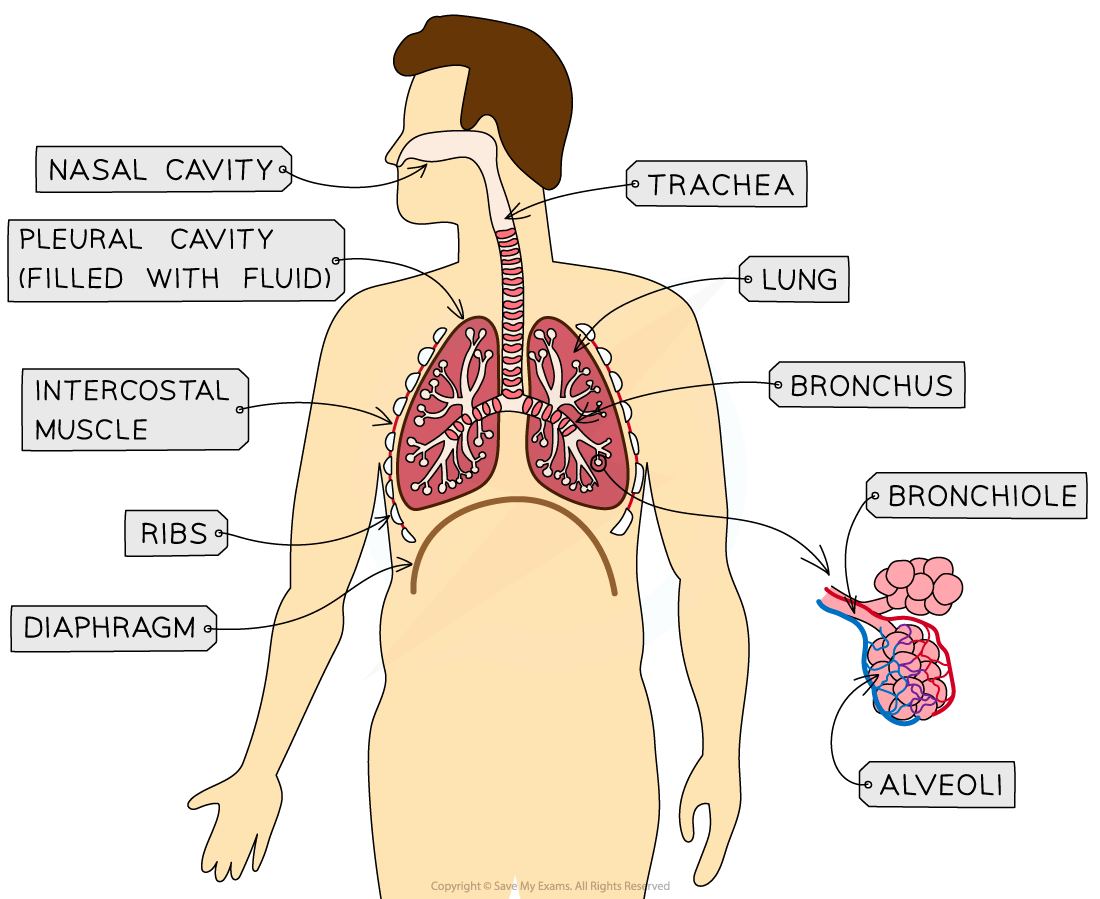
Human gas exchange occurs in the thorax. This system is the collection of organs and tissues that are present in the chest cavity. Now, let us see what structures are present in the human thorax.
- Trachea: This is the airway that extends from the mouth and nose to the bronchi. It is lined with mucus and secretes cilia and Goblet cells. The function of the cilia is to sweep away the dust and microorganisms from the lungs.
- Lungs: There are two lungs in the human body which are the main component of the respiratory system. Gas exchange takes place in the lungs.
- Bronchi: The left and right bronchi are located at the bottom of the trachea. Their structure resembles the trachea, however, they are narrower. The bronchi are further divided into bronchioles.
- Bronchioles: Bronchioles refer to narrow tubes that are typically less than 1mm. They transport air from the bronchi to the alveoli. Because of their narrow structure, they do not contain any supporting cartilage and hence they can collapse.
- Alveoli: They are the primary site of gas exchange in the lungs. They are small sacs with several structural adaptations to allow the efficient exchange of gas. The structural adaptations which support efficient gas exchange include thin walls and a larger surface area to volume ratio.
- Capillary Network: The capillary network includes an extensive network of capillaries that surrounds the alveoli and are an exchange surface between the lungs and blood. During the exchange of gases, the oxygen diffuses from the alveoli and into the capillaries. On the other hand, the carbon dioxide diffuses the other way and is exhaled.
Gas Exchange Tissues
The gas exchange tissues are explained below:
- Cartilage: It is a strong and flexible tissue present in different places around the body. One such place where the cartilage is present is in the rings along the trachea which are known as tracheal rings. These rings provide support to the trachea and ensure that it stays open. They enable it to move and flex during breathing.
- Ciliated Epithelium: It is a specialized tissue present along the trachea down to the bronchi. There are tiny projections of cilia in each cell that are responsible for sweeping dust, mucus, and bacteria upwards and away from the lungs and the epithelium itself.
- Goblet Cells: These cells are distributed throughout the ciliated epithelium in the trachea. These cells produce mucus that secretes viscous mucus to trap bacteria, dust, and other microorganisms. Due to the mucus secretion capability of the goblet cells, dust, bacteria and other microorganisms cannot reach the lungs. After that, the mucus is swept away along the cilia of the ciliated epithelium upwards and gets swallowed. The mucus along with any other microorganisms is then destroyed in the stomach by its acid.
- Alveoli: The alveoli have a lining of thin and squamous epithelium that enable gas exchange. The squamous epithelium creates a structure of the alveolar wall. Hence, it is extremely thin and permeable to enable easy diffusion of gases.
- Smooth Muscle: It is present throughout the walls of the bronchi and bronchioles. It assists in regulating the flow of air into the lungs through dilation when more air is required and constriction when less air is required.
- Network of capillaries: An extensive network of capillaries around each alveolus. The diffusion of carbon dioxide takes place out of the capillaries and into the alveoli to be exhaled. On the other hand, the diffusion of oxygen takes place the other way from the alveoli and into the capillaries to be transported around the body. The diameter of these capillaries is approximately 3-4 µm, which is sufficiently wider for a single red blood cell to move through at any one time. It ensures that enough time and opportunity are available for gas exchange to take place.
In the next section of the article, we will explain how to use the microscope to detect the structures present in the gas exchange system.
Using a Microscope to Recognize the Structures Present in the Human Gas Exchange System
Apparatus Needed
The main parts of an optical microscope are:
- The objective lenses
- The eyepiece lens
- The stage
- The coarse and fine focus
- The source of light
Other tools include:
- Forceps
- Coverslip
- Scissors
- Slides
- Scalpel
- Pipette
Procedure
Use a liquid specimen to prepare a slide in this way:
- Add some drops of the sample to the slide using a pipette
- Cover the smear/liquid with a coverslip and press it down gently to remove air bubbles
- Remember to wear gloves to ensure there is no cross-contamination of foreign cells
Use a solid specimen to prepare a slide in this way:
- Cut a small sample of tissue using scissors
- Cut or peel away an extremely thin layer of cells from the sample tissue to be placed on the slide by using forceps or a scalpel
- Treat some tissue samples with chemicals to kill or make the tissue rigid
- Place the coverslip gently on the top and press it down to remove any air bubbles present.
- A stain may be needed to create the visible structures, depending on the kind of tissue being examined.
- Be careful while using sharp objects and wear gloves so that the stain cannot dye your skin.
- While using an optical microscope, always start with the low-power objective lens. This is because you can find your desired object easily in the field of view. Moreover, it also prevents damage to the lens or coverslip if the stage is raised too high.
- Prevent the dehydration of the tissue by adding a drop of water to the specimen.
- To obtain a clearer image, switch to the lower power objective lens and try to use the coarse focus.
How to use a graticule to take measurements of cells?
- A graticule is a tiny disc that has an engraved ruler. It can be placed into the eyepiece of the microscope to play the role of the ruler in the field of view.
- Because the graticule has no fixed units, hence it should be calibrated for the objective lens that is in use. This is achieved by employing a scale engraved on a microscopic slide.
- By employing the two scales together, the number of micrometres that are equal to each graticule unit can be worked out
- Once this is known, the graticule can be employed as a ruler in the field of view.
Limitations
- In various specimen slides, the size of cells or tissue structures may appear inconsistent. The structures in cells are three-dimensional and various tissue samples would be cut at varying planes resulting in inconsistencies when observed on a 2D slide
- Since optical microscopes do not have the same magnification power as other types of microscopes, hence few structures cannot be observed.
- How specimens are treated while preparing the slides could change the structure of the cells.
Summarise with AI:
Did you like this article? Rate it!