In this article, we will discuss the structure of the human kidney, limited to the fibrous capsule, cortex, medulla, renal pelvis, ureter, branches of the renal artery, and renal vein. We will also Identify the parts of a nephron and its related blood vessels and structures like glomerulus, Bowman’s capsule, proximal convoluted tubule, a loop of Henle, distal convoluted tubule, and collecting duct in diagrams, photomicrographs, and electron micrographs. So, let us get started.


Anatomy of Kidneys
- Pairs of red-brown organs connected to the back of the abdominal cavity are known as kidneys. A thick protective layer of fat and connective tissue surrounds these kidneys.
- Renal arteries supply blood to the kidneys at arterial pressure which branches off the abdominal aorta.
- Through the renal vein, the blood exits the kidneys, into the inferior vena cava.
- The kidneys play a critical role in excretion, blood filtration, and osmoregulation.
In the next section of the article, we will discuss the functions of kidneys and their related structures.
The Function of Kidneys and Their Related Structures
The structures related to kidneys are:
- Renal artery: It transports oxygenated blood, i.e. the blood that contains salts and urea to the kidneys
- Renal vein: It transports deoxygenated blood, i.e. the blood that had urea and excess salts removed, away from the kidneys
- Kidney: It regulates the water content of the blood and helps in blood filtration
- Ureter: It transports urine from the kidneys to the bladder
- Bladder: Urine is temporarily stored in the bladder which is a muscular sac
- Urethra: This structure releases urine outside of the body
In the next section of the article, we will discuss the structure of human kidneys and nephrons in detail.
Kidneys
- Two kidneys present in the human body are responsible for carrying out the following two main functions:
- Kidneys act as an osmoregulatory organ as they regulate the water content of the blood which is critical for maintaining blood pressure.
- Kidneys act as excretory organs too because they excrete harmful waste products of metabolism such as urea and substances which are present in excess. Example of these substances includes salts
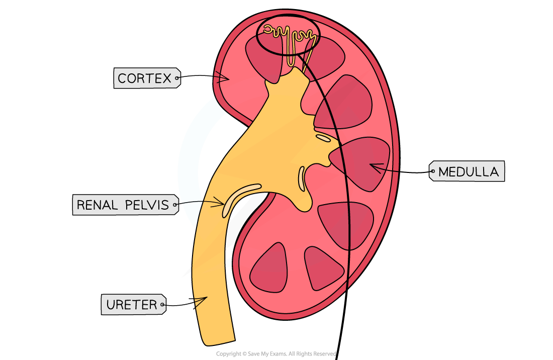
- A fairly tough outer layer referred to as a fibrous capsule surrounds the kidneys
- The kidney has three primary areas below the fibrous capsule:
- The cortex: The cortex is a dark outer layer that contains a high density of capillaries because it is the site where blood filtration takes place. It has not only a glomerulus but also Bowman’s capsule. Moreover, it also contains the proximal convoluted tubule and distal convoluted tubule of the nephrons.
- The medulla: The medulla is the lighter region within the cortex. It has the loop of Henle and collecting duct of nephrons
- The renal pelvis: The pelvis is the innermost structure of the kidney. It is where the ureter connects to the kidneys. It accumulates urine before it is passed down the bladder.
- Thousands of small tubes known as nephrons are present in each kidney
- These nephrons act as small filtering units
- The nephron, being the functional unit of the kidney is responsible for forming the urine
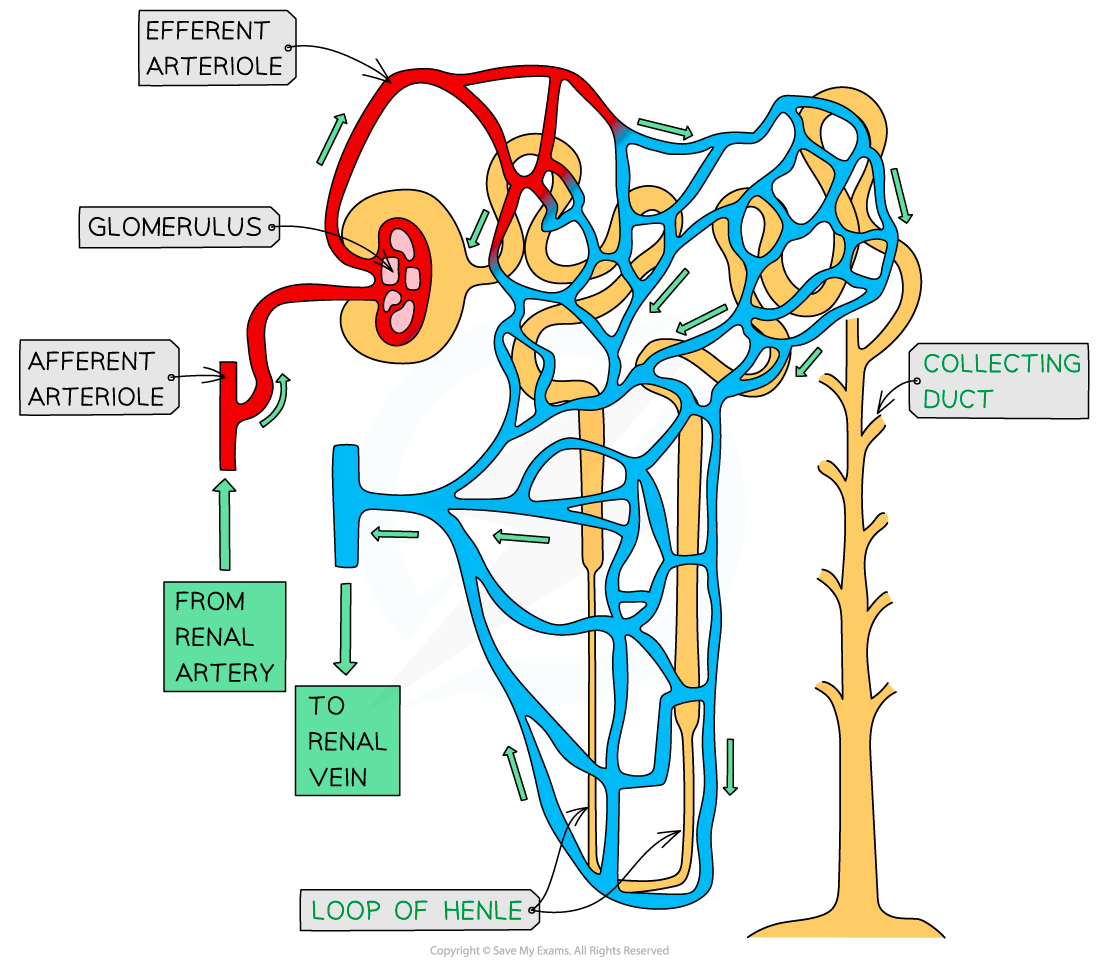
- A network of blood vessels is also connected with each nephron:
- A structure known as glomerulus is present within the Bowman’s capsule of each nephron
- An afferent arteriole supplies blood to each glomerulus. The afferent arteriole carries blood from the renal artery.
- The capillaries of the glomerulus join again to create an efferent arteriole
- After that, the blood flows from the efferent arteriole into a network of capillaries that closely run alongside the remaining nephron
- From these capillaries, the blood finally flows into the renal vein.
In the next section of the article, we will discuss nephrons and the structure of nephrons in more detail.
Nephrons
- Blood gets filtered in nephrons and most of the filtered material comes back into the blood
- It eliminates nitrogenous waste and balances water levels and mineral ions in the blood
- The length of each nephron is about 3 cm and there are 1.5 million nephrons in one kidney. This offers the body many kilometres for the reabsorption of water, glucose, salts, etc.
Now, let us discuss the structure of the nephron in detail.
Structure of Nephrons
A nephron contains the following structures:
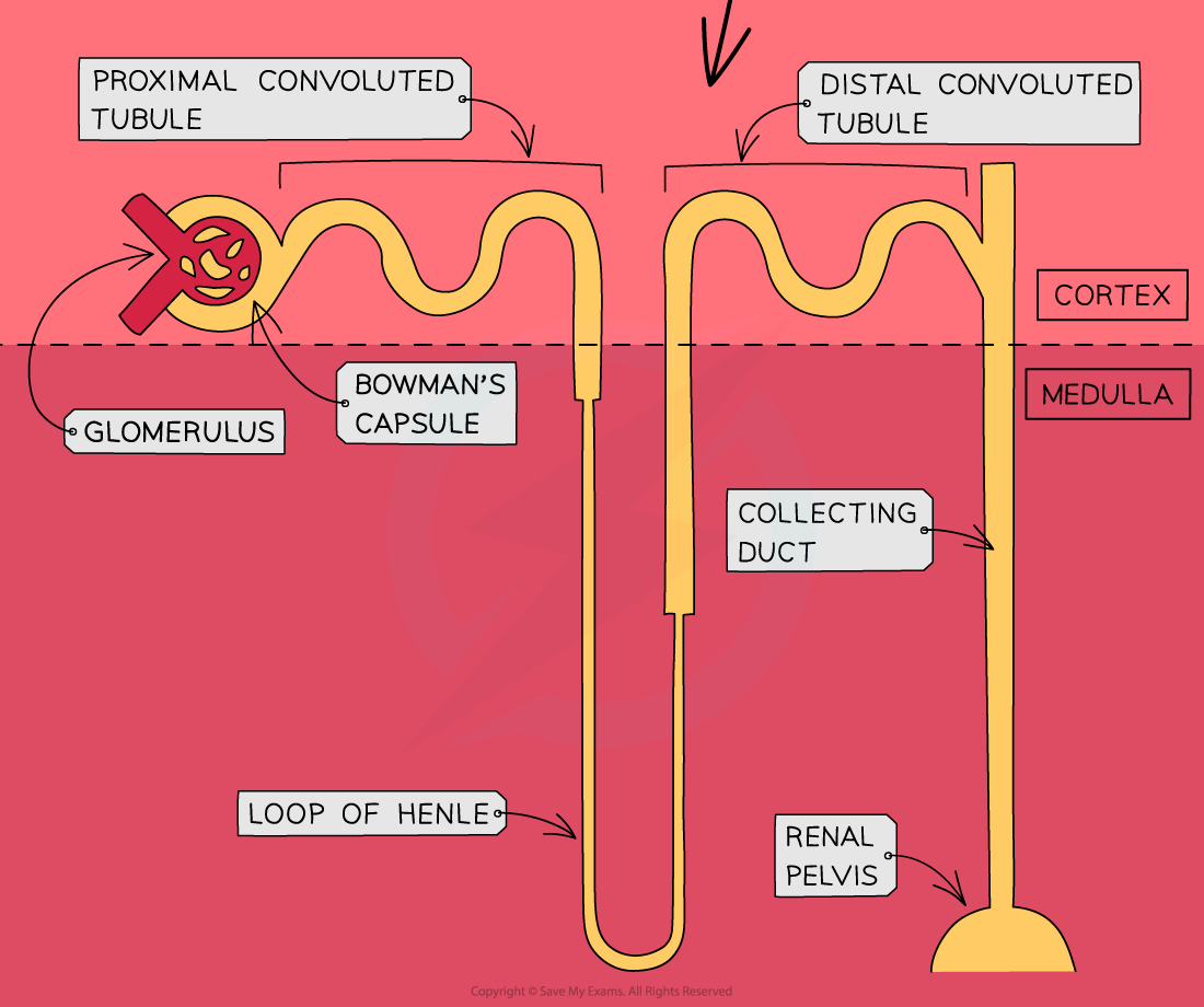
- Bowman’s Capsule: It is a cup-shaped structure that contains a glomerulus. It is the place where ultrafiltration occurs
- Glomerulus: It is the mesh of capillaries in which the pressure compels all solutes in the blood plasma to be forced through the walls of the capillaries. This entails ions, glucose, urea, amino acids, and water. Since proteins and erythrocytes are very large, hence they cannot pass through.
- Proximal Convoluted Tubule: It is the primary coiled region of the tubule. It is the place where products that are required in the blood such as glucose, amino acids, and ions get reabsorbed into the blood.
- Loop of Henle: It is a long loop of tubules that covers the cortex and medulla. It is employed to concentrate the urine. A salty environment is created in the medulla so that the water can osmose of water out of the nephron on the falling limb. Moreover, the impermeable rising limb enables salts to diffuse out, so that salty conditions are maintained.
- Distal Convoluted Tubule: It is the second coiled area of the tubule. It is the place where diffusion and osmosis of solutes take place for fine-tuning the water potential and pH of the blood. The permeability of the distal convoluted tubule is affected by the antidiuretic hormone (ADH).
- Collecting Duct: Urine moves through the collecting duct down to the pelvis. As ADH creates aquaporins to enable the exit of the excess water, more fine-tuning takes place.
Summarise with AI:
Did you like this article? Rate it!