In this article, we will discuss chromosome behaviour in mitosis. We will specifically explain the behaviour of chromosomes in plant and animal cells during the mitotic cell cycle and the related behaviour of the nuclear envelope, the cell surface membrane, and the spindle. We will also interpret photomicrographs, diagrams, and microscope slides of cells in different stages of the mitotic cell cycle and identify the main stages of mitosis.
Chromosomal behaviour refers to the routine movement of chromosomes during cell division. During mitosis, chromosomes that are replicated align themselves along the equator of the cell to create a metaphase plate. After that, sister chromatids are pulled apart towards the opposite poles of the cells. These overly complicated activities are carried out in a synchronized way. Different signals control not only these activities, but also the intra-cellular mitotic checkpoints.
Before discussing the chromosomal behaviour in detail, first, let us recall the structure of chromosomes.


Structure of Chromosomes
Chromosomes are composed of a single, extremely long condensed DNA molecule that is related to proteins in eukaryotic cells. The important proteins present are the large positively charged globular proteins known as histones. Their function is to organize and condense the DNA tightly so that it can fit into the nucleus. Enzymes are other proteins that are employed in the replication and repairing of DNA.
Chromatin refers to the strictly coiled combination of DNA and proteins. Both chromatids and chromosomes are composed of chromatin. DNA replicates during the interphase, i.e. the S phase to generate two similar strands of DNA known as chromatids. These strands are joined together by a narrow area referred to as centromere.
The two chromatids that create the dual structure of chromosomes are called sister chromatids. The sister chromatids must contain the same genes which means that they must be identical. This is because identical sister chromatids are key to cell division as one chromatid goes into one daughter cell and the other goes into the other daughter cell during mitosis. They do so to ensure that the daughter cells are genetically identical.
Each chromatid is composed of a very long, condensed DNA molecule. This DNA molecule is composed of a series of genes. The chromatids' ends in chromosomes are sealed with protective structures known as telomeres.
In the next section of the article, we will discuss the significance of mitosis.
Significance of Mitosis
Mitosis refers to the process of nuclear division which produces two genetically identical nuclei that are also genetically identical to the parent nucleus. The process of mitosis has huge biological significance as it is key to several biological processes. This process plays an important role in:
- Growth of multicellular organisms
- Asexual reproduction
- Replacement of cells and tissue repair
The behaviour of Chromosomes During the Mitotic Cycle
Now, let us discuss the behaviour of chromosomes during the mitotic cycle.
The process of mitosis is divided into the following four main stages:
- Prophase
- Metaphase
- Anaphase
- Telophase
The majority of organisms contain several chromosomes in the nuclei of their cells. For instance, humans have 46 chromosomes. Now, we will explain the behaviour of chromosomes in each of the four stages of the mitosis process.
Prophase
- The condensed chromosomes can be observed when stained.
- The chromosomes are made up of two identical chromatids known as sister chromatids. Each sister chromatid contains one DNA molecule. The centromere joins together the sister chromatids.
- The two centrosomes that are replicated in the
 phase right before the prophase travel towards the opposite ends of the nucleus, i.e. the opposite poles
 - Spindle fibres, i.e. protein microtubules start emerging from the centrosomes that are composed of two centrioles in animal cells
- The nuclear membrane (nuclear envelope) breaks down into tiny vesicles
Metaphase
- The centrosomes come to the opposite poles
- Spindle fibres, i.e. the protein microtubules continue to stretch from centrosomes
- At the equator of the spindle, i.e., the metaphase plate, the chromosomes line up in such a way that they are equidistant to the two centrosome poles
- Protein microtubules (spindle fibres) come to the chromosomes and attach to the centromeres
- Each sister chromatids joins with a spindle fibre that comes from the opposite poles
Anaphase
- The sister chromatids detach at the centromere which means that the centromere divides into two
- Spindle fibres start shortening
- Spindle fibres pull the detached sister chromatids, now known as chromosomes, to the opposite poles
Telophase
- Chromosomes reach the opposite poles and start to decondense
- Nuclear envelopes, i.e. the nuclear membranes start reforming around each set of chromosomes
- The spindle fibres disintegrate
In the next section of the article, we will interpret photomicrographs, diagrams, and microscope slides of cells in different stages of the mitotic cell cycle and identify the main stages of mitosis.
Mitosis in Root Tips (Observation and Drawing)
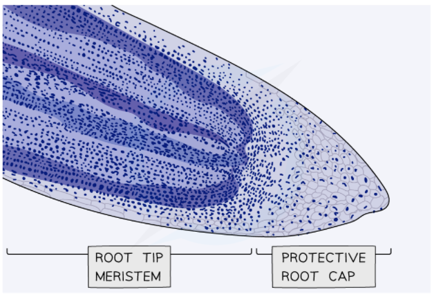
Meristems are the regions where growth in plants takes place. The root tip meristem can be employed to study mitosis. The root tip meristem can be present right before the protective root cap. There is a zone of cell division that has cells undergoing mitosis in the root tip meristem.
We can study pre-prepared slides of root tips. Besides this, we can also prepare temporary slides using the squash technique. In this technique, root tips are stained and squashed slightly to spread the cells out in a thin sheet so that the individual cells that are undergoing mitosis can be observed clearly.
Procedure
- The widely used root tips used in this experiment are the onion and garlic. Suspend the bulbs over water for one week or two, so that they can grow roots
- Remove the root tips and place them in an appropriate stain. For instance, warm acidified acetic orcein that stains the chromosomes deep purple.
- The stained root tip is squashed carefully on a glass slide by using a blunt tool such as the handle of a mounting needle
- You can observe and draw the cells undergoing mitosis as shown in the images below
- Add the annotations to the drawings to show various stages of mitosis.
The following micrograph shows the stained root tip:
Analysis of the Results
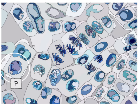
The following micrograph shows the cells undergoing prophase:
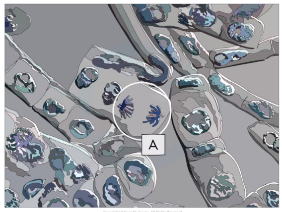
The following micrograph shows the cell undergoing the anaphase and metaphase:
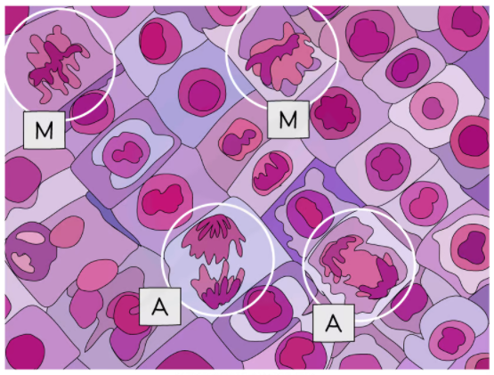
The following picture shows the micrograph of the cell undergoing metaphase and anaphase:
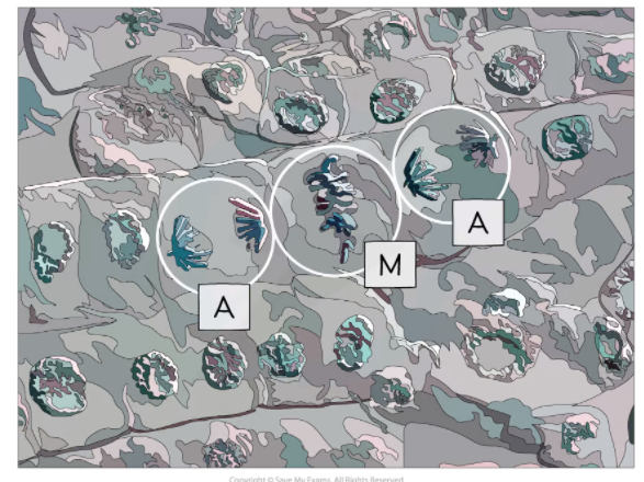
The following picture shows a micrograph of the cell undergoing anaphase:
Summarise with AI:
Did you like this article? Rate it!