In this article, we will discuss what roles neuromuscular junctions, the T-tubule system, and sarcoplasmic reticulum play in stimulating contraction in striated muscle. We will also explain the ultrastructure of striated muscle while referring to sarcomere structure using electron micrographs and diagrams. In the end, we will explain the sliding filament model of muscular contraction which includes the roles of troponin, tropomyosin, calcium ions, and ATP. So, let us get started.


Stimulating Contraction in Striated Muscle
- Striated muscle undergoes contraction which it receives an impulse from a motor neuron through the neuromuscular junction
- Due to the action potential, calcium ions diffuse into the neuron when an impulse that transfers along the axon of the motor neuron reaches the presynaptic membrane
- This triggers i.e. stimulates the vesicles which contain neurotransmitter acetylcholine (ACh) to fuse with the presynaptic membrane
- The released acetylcholine (ACh) diffuses across the neurotransmitter junction and binds to the receptor proteins present on the sarcolemma which is the surface membrane of the muscle fiber cell
- This triggers the opening of the ion channels in the sarcolemma, enabling the sodium ions to diffuse in
- As a result, the depolarization of sarcolemma takes place which generates an action potential that moves down the T-tubules towards the center of the muscle fiber
- Due to these action potentials, the voltage-gated calcium ion channel proteins in the sarcoplasmic reticulum membranes open
- The diffusion of calcium ions out of the sarcoplasmic reticulum and into the sarcoplasm that surrounds the myofibrils occurs
- The binding of calcium ions to troponin molecules stimulates a change in shape
- Consequently, troponin and tropomyosin proteins alter their position on the thin (actin) filaments
- On the actin molecules, the myosin-binding sites are exposed, and the process of muscle contraction referred to as the sliding filament model can now start
In the next section of the article, we will discuss the ultrastructure of striated muscle while referring to sarcomere structure using electron micrographs and diagrams.
Ultrastructure of Striated Muscle
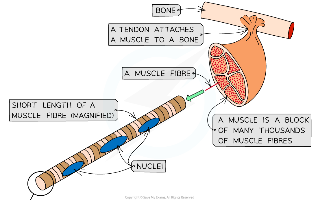
- Muscles in the body are made up of striated muscles that are attached to the skeleton.
- Striated muscle is composed of muscle fibers (a muscle fiber is a specialized cell-like unit)
- An organized arrangement of contractile proteins in the cytoplasm is present in each muscle fiber
- A cell surface membrane surrounds each muscle fiber
- Several nuclei are present in each muscle fiber, therefore we do not consider muscle fibers as cells
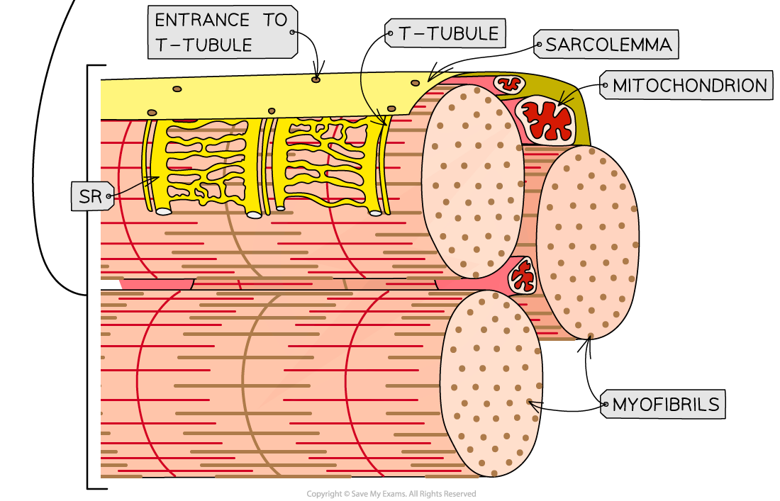
- Different names are given to various parts of the muscle fiber. These parts are equivalent to a normal cell. For instance:
- Cell surface membrane of the muscle fiber is known as the sarcolemma
- The cytoplasm of the muscle fiber is called the sarcolemma
- The endoplasmic reticulum of the muscle fiber is known as the sarcoplasmic reticulum (SR)
- Several deep tube-like projections that fold in from its outer surface are present in the sarcolemma. They run close to the SR and are referred to as transverse system tubules or T-tubules
- Mitochondria and myofibrils are present in the sarcoplasm
- The mitochondria undergo aerobic respiration to produce the ATP needed for muscle contraction
- Myofibrils refer to the bundles of actin and myosin filaments that slide past each other during muscle contraction
- Protein pumps are present in the membranes of the SR that are responsible for transporting calcium ions into the lumen of the SR
Myofibrils
- Myofibrils are present in the sarcoplasm
- Each myofibril is composed of two types of protein filament:
- Thick filaments are composed of myosin and thin filaments are composed of actin
- Both thick and thin filaments are arranged in a specific order to form different types of bands and line
Parts of Myofibrils and their Description
The various parts of the myofibrils are:
- H band: Thick myosin filaments are found only
- L band: Only thin actin filaments are found
- A band: It has regions where myosin filaments are present only and parts where myosin and actin filaments overlap
- M line: It is an attachment for myosin filaments
- Z line: It is an attachment for actin filaments
- Sarcomere: It refers to the section of myofibril between two Z lines
In the next section of the article, we will explain the sliding filament model of muscular contraction which includes the roles of troponin, tropomyosin, calcium ions, and ATP.
Sliding Filament Model of Muscular Contraction
In this section, we will explain the Structure of thick & thin filaments in a myofibril.
- Within a myofibril, the thick filaments are composed of myosin molecules
- Myosin molecules are fibrous protein molecules having a globular head
- The fibrous section of the myosin molecule anchors the molecules into the thick filament
- Several myosin molecules lie next to each other in a thick filament with their globular heads that point away from the M line
- The thin filaments inside the myofibril are composed of actin molecules
- Actin molecules are globular protein molecules
- Several actin molecules link together to create a chain
- The twisting of two actin chains results in the formation of a single thin filament
- A fibrous protein called tropomyosin is twisted around two actin chains
- Another protein referred to as troponin attaches to the actin chains at regular intervals
Contraction of Muscles – the Sliding Filament Model
The movement of muscles is caused by contraction. While muscle contracts, the sarcomeres inside the myofibrils shorten because the Z discs are pulled closer together. It is referred to as the sliding filament model of muscle contraction and takes place through the process described below:
- At the neuromuscular junction, an action potential arrives
- From the sarcoplasmic reticulum (SR), calcium ions are released which bind to the troponin molecules, triggering them to alter their shape
- Subsequently, tropomyosin and troponin proteins alter their position on the actin, i.e. thin filaments
- On the actin molecules, the myosin-binding sites are exposed, and the globular heads of the myosin molecules bind with these sites to create cross-bridges between the two types of filament
- The myosin heads move and pull the actin (thin) filaments towards the sarcomere’s center, resulting in the small distance contraction of the muscle
- At the myosin heads, the hydrolysis of ATP takes place which provides the needed energy for the myosin heads to release the actin filaments
- The movement of myosin heads to their original position occurs along with their binding to new binding sites on the actin filaments, that are near the Z disc
- Once again, the movement of myosin heads occurs which pulls the actin filaments even closer to the center of the sarcomere. Consequently, the shortening of sarcomere occurs along with the pulling of the Z discs closer together
- Once more, the myosin heads hydrolyze ATP to detach again
- The process repeats until the full contraction of the muscle, until and unless the troponin and tropomyosin are not blocking the myosin-binding sites and the muscle has an ATP supply
Summarise with AI:
Did you like this article? Rate it!